Flowers

Above and below: Passiflora, the passion flower. For many years I would say that the passion flower was my favourite flower, but to be honest I don't think I have a favorite (it was the tulip once, then the lupin, then the snapdragon, then the bluebell ...). The more I learn about different plants the more I appreciate them all! Indeed, one of the great fascinations about Nature is the diversity of forms that it generates - variety is the spice of life, as they say!
True flowers only occur in the Angiosperms, though the term is sometimes loosely applied to reproductive structures in Bryophytes and Gymnosperms. Flowers are modified shoots in which the sterile and fertile reproductive organs are borne on an axis (the receptacle). This modified shoot exhibits determinate growth meaning that it ceases growth once the flower is developed (the floral meristem ceases activity after all the floral parts have been produced). Individual parts of the flower may continue to show growth, as the small growth changes which bring about opening and closing of flowers in response to temperature and light (nyctinasty).
The parts are arranged in whorls rather than in a spiral or helix. Cohesion, that is fusion or joining together of members of a whorl is frequent and the whorl then grows as a unit. For example, petals may cohere to form a flower-tube (coralla or petal tube). Adnation is the fusion of one whorl to another, in which case two or more whorls grow as a unit. For example, the fusion of anther filaments to petals.
Flowers are the sexual reproductive organs of the flowering plants (angiosperms) and consist of sterile and fertile
reproductive organs borne on the swollen end of a shoot axis (receptacle). The flower is a modified leafy (vegetative) shoot. The leaf-bearing nodes of the shoot now bear structures derived from leaves and the internodes between these nodes are greatly shortened to bring several whorls of these modified leaves together. Like all shoots, flowering shoots grow from buds in the axils of leaves (axillary buds) on the parent shoot. These buds typically produce green leafy shoots, but when the time for flowering arrives some of these buds switch to a different developmental program and put out an inflorescence (flowering shoot) bearing one or more flowers, or flowers may simply develop at the ends of vegetative shoots. The leaf accompanying this bud may become modified and is known as a bract. The bract now sits at the base of the inflorescence. The inflorescence puts out one or more flowers, each borne on its own stalk or pedicel and each deriving from a terminal flower bud. Once a bud switches to become a flower the shoot ends there - no more branches will be produced from this shoot tip and it becomes destined to become a flower and the meristem, or growing tip, ceases activity once all the flower parts have been produced.
Sepals and Petals
The various organs of the flower are arranged in whorls that encircle the stem, rather than in helices or spirals as are most leaves. The lowest whorl of leaf-like structures to make up the flower is called the calyx and is made up of leaf-like sepals. Sepals are often green and photosynthetic, like ordinary leaves, but differing in shape and with a more compact internal structure. In this case the calyx is referred to as sepaloid. In others they turn into hard scales to protect the developing bud and they may be shed once the flower opens. Sepals may remain attached to the flower and fall off when the flower withers or they may remain and form part of the fruit, possibly contributing to the flesh of the fruit. In the passion flower above, the sepals are five in number and look much like the petals, but are greener in color and end in hook-like awns. A calyx comprising sepals that resemble petals is called petaloid.
Sepals and petals resemble leaves in structure: they consist of parenchyma, have a more or less branched vascular system and an epidermis. Crystal-containing cells, laticifers, tannin cells and other idioblasts (cells containing special substances) may be present. Young petals may contain starch. Green sepals contain chloroplasts but rarely have differentiated palisade and spongy mesophylls. Petals contain pigments in chromoplasts (carotenoids) and in the cell sap (flavonoids, e.g. anthocyanins). Some of these pigments may reflect UV to act as guides to pollinating insects. Epidermal cells of petals often contain volatile fragrant oils. The epidermis of both sepals and petals may have stomata and trichomes (hairs, which may be glandular and secrete various substances).

The
next whorl of 'leaves' along the shoot develop into petals that together constitute
the corolla- they are white in our
passion flower. The internal structure of petals is much like that
of green leaves, except that they are modified to perform less (if
any) photosynthesis and instead their plastids may store pigments to
give the petals color. In green leaves these plastids develop into
chloroplasts which contain green chlorophyll, giving the leaf its
green color. Chlorophyll absorbs light and harvests energy from the
light to make sugars from carbon dioxide gas from the air and water.
From these sugars the plant will make all the building materials it
needs to make its body - fats, proteins, nucleic acids, long-chain
carbohydrates, etc. The roots supply both the necessary water (in
most plants) and mineral ions from the soil.
Chloroplasts
also contain yellow-orange-red pigments called carotenoids. Usually these pigments
are masked by the green chlorophyll (except in plants with reddish
leaves like copper beech) but become visible in autumn when
chlorophyll has been broken down and resorbed by the plant, leaving
behind the carotenoids to give the leaves their splendid autumn
colors. Pigments may also be stored inside the plant cell vacuole, principally
flavonoids like anthocyanins which are red, purple or
blue depending upon the pH (acidity) of the cell sap within the
vacuole. Anthocyanins also add to the autumn colors of leaves.
Many
petals also have a special microstructure to their surface tissue
which comprises an epidermis (that is a sheet of covering cells) of
conical cells bearing ridges. These ridges help to focus the light
and enhance the petal's color. The petals of some flowers remain
green and photosynthetic. These flowers tend to be wind pollinated
and are usually small. Plants with large, showy, colorful flowers
are advertising for animals to come and pollinate them. Insects,
birds, bats and other animals may act as pollinators. Some flowers
provide only excess pollen as food, with which to reward their
animal pollinators, but others, like the passion flower, possess nectaries that secrete a sugary nectar
that is often fine-tuned to meet the dietary needs of the flower's
favorite pollinator. The epidermal cells of the petals may secrete fragrant oils to help lure their animal
pollinators.
In the passion flower some of the petals are modified into coronal
filaments, forming the spectacularly colorful 'crown of thorns'.
These colorful flowers are easy for the animal to spot at a distance
and the arrangement of petals and their colors guide the animal to
the food reward of pollen and/or nectar. It is in a sense by chance
that many insect-pollinated flowers are colorful to our eyes, since
although some insects, like bees, have good color vision they are
also able to see ultraviolet light, which gives them a very
different perception of color, and to them a flower looks very
different as they see colors differently. In ultraviolet many
insect-pollinated flowers bear additional markings, often stripes
that guide the insect straight to the nectar, rather as markings
guide a taxiing plane on a runway. I say, 'in a sense' because most
mammals can not see colour at all. Primates are a rare exception,
they have color vision probably to help them locate fruit in the
canopy. Since fruit possess many similar pigments and colors as
flowers, we can see both.
The calyx of sepals and the corolla of petals are the sterile parts
of the flower and together they form the perianth. Sometimes the sepals and
petals are very alike and hard to distinguish, in this case they are
called tepals.

Finally we come to the fertile organs of the flower - the male stamens (microsporophylls) that together form the
androecium and the female carpels (megasporophylls) that form the gynoecium.
Many flowers have both male and female organs and are called perfect flowers. In some species flowers may have only the male parts (staminate flowers) or the female parts (carpellate or pistillate flowers) and these are called imperfect flowers. In species with imperfect flowers, male and female flowers may occur on the same branch, different branches or on separate plants. In the latter case the plant has distinct male and female individuals and is said to be dioecious. Many plants, however, are far more confused and may contain male, female and individuals that are hermaphrodite to a variable degree.
The stamens (also called microsporophylls, which literally means leaves producing microspores) were also derived from leaves over the course of evolution. Indeed, the stamens of some plants are still leaf-like and bear sporangia (spore producing organs) underneath, rather like ferns and other non-flowering plants that produce spores from sporangia underneath fertile fronds. The fossil record tells us that flowering plants appeared quite late in evolution (and genetic analysis reveals that their dramatic speciation is in large part due to their tendency to hybridise) and some flowering plants still bear such 'primitive' or archaic characteristics. The stamens of most flowers, however, comprise an anther head borne on a slender stalk or filament. The anther produces pollen (microspores) in (usually four) pollen sacs (microsporangia) within it. The pollen contain the male sperm and when ripe the anthers dry and rupture (dehisce, dehiscence) to release their spores. The anthers may dehisce by splitting to form a longitudinal slit (as in cotton) or a transverse slit (as in basil) or a single pore called the stomium or a number of pores (e.g. potato) or they split into a number of valves as in bay leaf. The layer of cells in the anther wall may contain strips that twist upon drying to bring about rupture.
The encasement of sperm in pollen is an adaptation to life on land as the tough casing of the pollen wall resists dry conditions and allows the sperm to be transported to a female plant by animal or wind, over many miles if need be. Moss, ferns and cycads and other archaic lineages are less well adapted to life on land and still produce naked sperm (as their algal ancestors did) that require a film of moisture in which to swim.
The female carpels may be separate (an apocarpous flower) but are often fused together into a single structure (a syncarpous flower). Sometimes the word pistil is used to describe a single carpel in an apocarpous flower or the single compound carpel of a syncarpous flower. The carpel is a modified folded leaf rolled up with its margins more or less fused together. The final structure has one or more internal cavities called locules, in which future embryos may develop. The style is the the upper elongated part of the pistil and is absent in some flowers. The style ends in the stigma, usually a bulbous structure. The remainder of the carpel is the ovary, or main body of the carpel. The stigma is the receptive region to which pollen adheres prior to fertilization. The pollen grain then grows a pollen tube down through the style to reach the ovary and then into an ovule ('little egg'), attached by a placenta to the ovary wall inside one of the locules and then it discharges the sperm through a pore in the ovule, called the micropore or micropyle. The sperm enter the embryo sac (which is derived from a megaspore) within the ovule, which contains the female egg cell (ovum) and a few other (haploid) cells. Only pollen from the right species is accepted and even then pollen from the same plant may be rejected, thereby favoring cross-pollination and outbreeding which keeps the genetic stock strong by preventing inbreeding.
Flowers have various other tricks to ensure cross-pollination, or to prevent self-pollination. One of these is protandry, in which the male organs develop before the female organs on the same flower. In the passion flower, the styles are erect when the flower opens initially, holding the stigmas well out of the way of the anthers and any insect visitors that may brush past the anther, collecting pollen, and then brush the stigma of the same flower. As the anthers become exhausted of pollen, the styles bend down, finally bringing the stigmas to the same level as the anthers. Now when an insect visits the nectaries there is no pollen left to collect, but it can easily brush the stigmas and deposit pollen from another plant.
The passion flower is what we call an actinomorphic flower, meaning that it has 'circular' symmetry (strictly radial symmetry). Some flowers, like the white deadnettle, Lamium alba, have zygomorphic flowers which have only a single plane of symmetry and distinct left and right halves and a distinct top and bottom (they have bilateral symmetry, as do human beings!). These are evolved from actinomorphic flowers by assymetrical fusion of the petals into a floral tube (the corolla). This restricts access to the nectar at the base of the floral tube to those pollinators that can reach it. This exclusivity evolves when a plant finds certain pollinators extremely reliable and so it rewards them by saving its nectar by not giving it to less reliable pollinators.
The white deadnettle (White Archangel, Lamium album) is also protandrous. In the male stage, the style has not yet elongated far enough to position the stigma near the entrance, but the anthers are in position and visiting animals will brush against them to collect pollen. In the female stage, the anthers are spent and the style has elongated to position the stigma right at the entrance to the flower, where visiting animals can easily brush against it and deposit pollen.


Above:
a half-flower diagram of Lamium
album in
the female stage (top) and earlier male stage (bottom left). Bottom
right is the floral diagram and floral formula. In the floral
diagram, the topmost small circle (sometimes drawn with a cross, +,
inside it) indicates the position of the axis of the flower
and tells us that we are looking straight down the end of the
receptacle, that is we are viewing the flower face-on. The black arc
beneath shows us which side the bract sits on, at the base of the
flower stalk. Next we have the outermost whorl of five sepals, drawn
bridged together to show that they are joined together (as they are
toward the base of the flower). When members of the same whorl join
together we say that they cohere and we have cohesion of the parts.
Inside the sepals are the five petals which have the four stamens
attached to them inside. The petals are coherent or joined to one
another. The fusion of members of different whorls, such as the
stamens to the petals, is called adnation, they are adnated.
Innermost are the two carpels, fused together into a single pistil
with four chambers or locules.
The ovary can be described as either inferior or superior. In an inferior ovary (epigyny, epigynous ovary)
the sepals, petals and stamens are borne on the receptacle above the
ovary - a more advanced condition than the superior ovary (hypogyny,
hypogynous ovary) in which the sepals, petals and stamens occur
below the ovary. In perigyny, an extension above the
receptacle resembling a cup (receptacular or appendicular floral
tube) bears the sepals, petals and stamens.
Finally, although many flowers are hermaphroditic many are also imperfect flowers, meaning they are
unisexual, lacking either the gynoecium (staminate
flowers)
or the androecium (carpellate
or pistillate flowers).
Sometimes both male and female parts are present, but one is
non-functional, in which case the flower can be described as functionally pistillate
or staminate.
The floral
formula
describes this flower to us - the dagger symbol tells us that the
flower is zygomorphic. K represents the sepals of the calyx, and we
see that there are five of them. The C is the corolla, which is made
up of five petals as indicated, but enclosed in brackets () to tell
us that the petals are fused together into a floral tube. A
represents the androecium, containing 4 members. The square brackets
[] around the corolla (C) and androecium (A) tell us that these two
whorls are adnate (fused to each other). Finally we have the
gynoecium (G) of two carpels fused together.
The pictures below (click to enlarge) show the snapdragon, another
zygomorphic flower which is also protandrous.


The
Carpel
The
female part of the flower consists of one or more carpels. Often the
word pistil is often used interchangeably with carpel, but a
pistil also refers to several carpels fused together. The gynoecium
is the total female system of the flower, whether consisting of a
single carpel, several separate carpels or a pistil of fused
carpels. The diagram below shows the structure of a typical
angiosperm carpel. The ovary is the main body of the
carpel and contains the ovule. The style is an extension of the
ovary wall that holds aloft the stigma, which is a surface that
is sticky to pollen and acts to receive and trap pollen grains. The
ovule is attached to the ovary wall via a stalk, called the funiculus, which connects to a
region of the ovary wall called the placenta. Vascular tissue (often
a single vessel) passes from the placenta along the funiculus.


The diagram below shows the structure of a typical angiosperm carpel. The ovary is the main body of the carpel and contains the ovule. The style is an extension of the ovary wall that holds aloft the stigma, which is a surface that is sticky to pollen and acts to receive and trap pollen grains. The ovule is attached to the ovary wall via a stalk, called the funiculus, which connects to a region of the ovary wall called the placenta. Vascular tissue (often a single vessel) passes from the placenta along the funiculus.
The
ovule develops as a protuberance projecting from the ovary wall in the region of the placenta, a mass of cells
hemispherical folds grow around it (around the outer cells or
nucellus), extending from the epidermis, by cell division, lining
the inside of the ovary wall and which grow to enclose the ovule in
almost complete spheres, called integuments. The integuments do not
close over the ovule completely, but maintain a small opening called
the micropyle. Since they are extensions
of the ovary epidermis, the integuments are each lined (on the
outside and inside) by cuticle.
Inside the developing ovule, the outer cell layers are vegetative
and form the nucellus. Inside the nucellus is
the
sporogenous region. The chalaza is the region of tissue
where funiculus, integuments and nucellus meet. The sporogenous
region begins as a large archesporial
cell,
which gives rise to a megaspore
mother cell
(MMC) either by direct differentiation or by undergoing a mitotic
division (see mitosis) into two daughter cells, a perietal cell and
the MMC. The MMC undergoes meiosis to produce four haploid megaspores (enclosed in secreted
callose walls which later dissolve). In most angiosperms, 3 of the 4
megaspores degenerate, but the remaining megaspore undergoes 3
successive mitotic divisions, doubling each time, to produce 8
haploid nuclei. These nuclei form the embryo
sac,
which is the female
gametophyte
(gamete producing 'plant').

The
eight haploid nuclei of the gametophyte are enclosed in 7 cells.
Three haploid cells form at the pole of the embryo sac opposite
furthest from the micropyle, these are the 3
haploid antipodal cells.
At the micropylar end, nearest the micropyle, three haploid cells
are situated, two
haploid synergids
in front of and either side of the single haploid
ovum or
egg cell; together these three cells are called the egg apparatus. These six cells are
surrounded by the large central
cell,
which has the two remaining haploid polar
nuclei
inside it (these two nuclei may fuse to form a single diploid
nucleus, called the secondary endosperm nucleus, in some species).
It has been shown that the integuments protect the embryo sac and
developing embryo and prevent it from being crushed as the ovule
develops, by taking up compressive stresses. The nucellus nourishes
the developing embryo sac and embryo and may be partially or
completely absorbed by contact with the embryo sac in which case it
appears to takes over the nutritive role. The layer of cells
immediately enclosing the embryo sac and forming its wall may
develop wall ingrowths as may the central cell (increasing the
surface area of their cell membranes, a characteristic that suggests
they are transporting materials into or out of the embryo sac).
Nutrients are transported into the embryo sac and central cell from
surrounding tissues supplied by vascular tissue through the placenta
and funiculus.
This type of embryo sac development is the most common in
angiosperms and is well-studied in Polygonum and is referred to as
monosporic development, since only one of the four megaspores
produces the embryo sac. In other types, development may be bisporic
(requiring 2 of the 4 megaspores) or tetrasporic (requiring all 4
megaspores). In these types of development not all the 8 nuclei
produced need be haploid (though the ovum always is) and triploid
(3n) or even tetraploid (4n) nuclei may result.
The synergids have incomplete cell walls, covering the two-thirds of
the cells that face the micropyle, leaving the naked cell membrane
to contact the central cell behind. These cell walls develop deep
finger-like invaginations, forming the filiform
apparatus.
The egg cell has a cell wall that also tends to be incomplete at the
chalazal end (exposing naked cell membrane for fusion with a sperm
cell at fertilization). In grasses the antipodal cells divide
further by mitosis, producing a many-celled short-lived tissue which
develops wall ingrowths where they border the nucellus, suggesting
that they may serve some role in the loading of nutrients into the
embryo sac.

Alternation
of Generations
Plants
exhibit an alternation of generations. In mammals the diploid adults
(diploid means that they have 2 copies of each chromosome (2n) one
from the father and one from the mother) produce haploid gametes,
eggs and sperm (haploid means that only one copy of each chromosome
is present) which fuse at fertilisation to form a diploid
single-celled zygote which develops (by mitosis) into the embryo. In
plants, reproduction is a more complicated affair. Plants alternate
between separate diploid
sporophyte
plants and haploid
gametophyte
plants. The sporophytes produce spores that give rise to the
gametophytes, which produces spores that become the gametes. In
ferns, these two generations are physically separate plants, with
the sporophyte being the dominant plant we know and the gametophyte
small and easily overlooked, in mosses and liverworts, the
gametophyte is the dominant plant and the sporophyte grows almost
parasitically from it. In angiosperms, the sporophyte forms the
dominant plant body and the gametophyte, not really now a separate
plant at all, forms a groups of cells firmly attached to the
sporophyte body. The female gametophyte is the embryo
sac, the
male gametophyte is the pollen
grain.
Anthers
and Pollen grains
The
stamen is a microsporophyll - a modified leaf that
produces microsopores, and consists of a
filament (stalk) bearing the anther. The anther contains pollen
sacs, which are the microsporangia in which the microspores are
produced and are made up of sporogenous tissue and surrounding wall
layers. For example, the anther of cotton (Gossypium
arboreum)
is bilobed (has two lobes) and is bisporangiate (has two
microsporangia).
The developing anther consists of an outer cell layer, the protoderm
(which becomes the epidermis) and an underlying (hypodermal) cell
layer of archesporial
cells.
The archesporial cells undergo periclinal mitotic divisions
(periclinal means parallel to the surface of the anther) so that
this single layer of cells becomes two cell layers, the outer or primary parietal (wall)
layer
and the primary sporogenous
cell layer.
The outer parietal layer undergoes another set of periclinal cell
divisions, producing two new cell layers, the secondary
parietal layers.
The outer secondary parietal layer undergoes a third periclinal
division to produce two tertiary
parietal layers.
Thus at this stage there are three parietal layers (the inner
secondary parietal and the middle and outer tertiary parietal cell
layers) and the inner sporogenous cell layer. The sporogenous cells
enlarge and transform directly (or by mitosis) into microspore mother cells (SMCs). Now, in total the
anther wall has 4 cell layers: the outer epidermis, an underlying
cell layer (the outer tertiary parietal layer) which becomes the endothecium, a middle cell layer (the
inner tertiary parietal layer) and the innermost wall layer or tapetum
(the inner secondary parietal layer, which also incorporates some
parenchyma cells from the central tissues of the anther). (In some
species there are two middle layers, as the inner secondary parietal
layer may also divide, making 5 layers in total). The middle layer
degenerates and becomes crushed between the tapetum and endothecium
and is absorbed by these cells, leaving 3 cell layers outside the
sporogenous tissue in a mature anther. Inside this is the layer of
spore mother cells and then the parenchyma connective tissue that
fills the centre of the anther. The endothecium (just beneath the
epidermis) develops secondary cell wall thickenings over its radial
walls and its inner most tangential wall (tangential = parallel to
the surface of the anther, i.e. the back wall), except in the region
of the stomium, which is the region where
the anther will open when releasing pollen.
The miscrospore mother cells (also called pollen mother cells or
microsporocytes) undergo meiosis to produce 4 haploid microspores (pollen grains in their
early stage with one nucleus each). At first, the SMCs are compacted
together and joined by plasmodesmata. During meiosis, callose walls
are deposited around them, the pre-existing cellulose cell walls
disintegrate, and the cells round-off, remaining connected to one
another by wide cytoplasmic bridges that traverse channels (of about
1.5 micrometre diameter) in the callose. These cytoplasmic
connections allows the SMCs to communicate rapidly with one-another,
ensuring that they develop in synchrony. (In Gymnosperms, including
conifers, there are no such connections and the SMCs develop
asynchronously). The connections disappear before the completion of
meiosis, so that each SMC gives rise to 4 isolated cells (a tetrad)
often in a tetrahedral arrangement. The tapetum (the innermost
parietal layer) develops in synchrony with the SMCs and its cells
become multinucleate or polyploid (they make multiple copies of each
chromosome) increasing the numbers of copies of each gene that they
have, allowing more rapid synthesis of proteins and they provide
nutrition to the developing spores. There are two types of tapetum.
Type 1 is glandular/secretory and lyses (they burst as their cell
walls rupture) after meiosis to deposit lipid-rich material, called
tryphine, over the maturing pollen
grains. Type 2 lose their cell walls and becom amoeboidal, fusing
together into a multinucleate plasmodium whose cell processes
protrude amongst the developing pollen and which dehydrates before anthesis (the time of complete
flower development and opening) to deposit tryphine over the
maturing pollen. The end result is essentially the same in both
cases.
The microsporocytes each become enveloped in a complex cell wall
with an outermost rigid layer called the exine and an inner flexible
layer called the nexine (a double layer) overlying the cell
membrane. There are pores in the exine, where the nexine thickens,
with one pore being usual in monocotyledons, three in most
dicotyledons. These pores may be circular or elongated slits. The
exine contains sporopollenin, a very tough and resistant polymer and
sometimes also silicon for extra hardness 9one of the few known uses
of silicon in plants). The exine is more or less sculptured. In
wind-pollinated flowers, the pollen grains are usually small and
smooth for easy transport, but in animal pollinated flowers they can
be larger (and so contain more food reserves) and are spikier,
allowing them to stick to animal bodies better. The nexine contains
pectin and cellulose. The exine pores allow the pollen tube to
emerge during pollen grain germination.
Before the pollen is shed, each grain originally containing a single
microspore nucleus, undergoes mitosis to produce two haploid nuclei,
a vegetative
nucleus (VN) and
a generative
nucleus (GN).
The generative nucleus undergoes a second mitotic division, either
before or after the pollen is shed, to produce two
sperm cells.
The sperm cells are spindle-shaped (microtubules form rod-like
bundles inside the cells to maintain their shape), lack cell walls,
and are joined together head-to-tail and surrounded by the
vegetative cell (thus each sperm cell is enclosed in a double cell
membrane, the inner cell membrane being its own and the outer
belonging to the vegetative cell).

Above: pollen grains of winter Jasmine.
Below: cross-sections through stamens of Chrsyanthemum, showing the filament and the two three apparent cell layers forming the anther walls and containing tetrads (groups of 4).


Below: more stamens of Chrysanthemum, on the right is a close up view of a section through the filament - click images to enlarge; note that the pollen grains are still maturing and are arranged in groups of four (tetrads):


Below: more Chrysanthem floral parts, left - petal in transverse section; middle - sepals in transverse section; right - carpel in transverse section; note that many of the parenchyma cells in the anthers, petals and sepals, are loaded with dense material which will be the pigments of the flower to give it its colour which is so attractive to pollinators:


Below: mature pollen grains of Chrysanthemum in section. Note that the pollen grains are now separated from their tetrads (by dissolution of the callose walls which bound them together) and that the wall of each pollen grain is made up of two main layers - the outer exine, which is sculpted, in this case into columns called pili (singular 'pilum') and the inner intine. The exine contains a very resistant material called sporopollenin, which is a polymer derived from carotenoids and carotenoid esters. The smoother intine contains pectin and cellulose. The pollen grains of some plants also incorporate silica, which adds to the resistance of the walls to biological decay and weathering. There are circular or elongated regions of the exine which are thinner and it is through one of these regions that the pollen tube will emerge. One or two such regions are visible in one of the pollen grains in the middle image. Monocotyledons usually have one such region, dicotyledons three.


1. Pollen attaches to the stigma of a receptive carpel, this is pollination.
2. Proteins on the pollen grain and stigma may determine compatibility in those species favouring cross-pollination, so that only pollen from another plant will germinate.
3. The pollen grain germinates - it grows an extension of the vegetative cell, called the pollen tube, through one of the pollen grain exine pores/apertures.
4. The VN and the two connected sperm cells (or GN if it is yet to divide) travel down the pollen tube as a single unit, with the cell organelles accompanying them. This movement is driven primarily by actin and myosin filaments of the cytoskeleton. The pollen tube grows near its tip and deposits a cell wall (of carbohydrate polymers) as it does so. The older basal regions of the pollen tube may be closed off by callose plugs behind the nuclei descending along it.
5. The pollen tube grows at about 1-2 mm/h, making its way through the style tissues and into the space around the ovule, following the ovule along the integument surface and entering via the micropyle, or sometimes working its way along the funiculus to the chalaza. If the pollen tube needs to cross any remaining nucellus, then the nucellar cells in its path disintegrate to provide easy passage.
Possible paths of pollen tube growth shown in red. Once one pollen tube reaches the synergid, degeneration of the synergid removes the attractive signal and other germinating pollen tubes cease development, so that only one pollen grain achieves fertilisation.

6. The synergid cells attract the pollen tube (by a mechanism that is not yet understood), one more so than the other. The pollen tube enters this synergid cell through its filiform apparatus, after which the synergid degenerates (the other synergid degenerates also, though often at a later time). The pollen tube tip opens inside the synergid and the two sperm cells emerge, separate and become tightly coiled. The VN also emerges and degenerates, along with the synergid nucleus, forming X-bodies.
7. The two sperms are directed by the synergid cell (apparently by its actin cytoskeleton), one is led to the ovum, the other to the central cell. the synergid cell membrane dissolves. The first sperm fuses with the ovum and enters it, after which the sperm and egg nuclei fuse to produce the diploid zygote. The second sperm fuses with the two polar nuclei (or the diploid endosperm nucleus formed by their prior fusion) to form a triploid fusion nucleus or primary endosperm nucleus. This peculiar double fertilisation is a characteristic of angiosperms.
8. The ovule develops into the seed, the integuments forming the seed coat and the ovary forming the fruit. The zygote develops into the plant embryo, whilst the primary endosperm nucleus gives rise to endosperm tissue which nourishes the developing embryo/seedling after seed germination.
Floral
Formulae
A
floral formula is a code indicating the number of flower parts,
whorls, the attachment of parts and the nature of the gynoecium.
E.g. Lamium
album
(white dead nettle):
% K(5) [C(5) A4] G(2)
The % indicates a zygomorphic flower, i.e. a flower with
bilateral symmetry, a dagger cross symbol is often also used.
Various symbols, such as a cross in a circle (we may use + as it
is easier to type!) indicates an actinomorphic flower, that is one
with radial symmetry. A @ is sometimes used to indicate spiral and
not whorled parts, K = calyx, C = corolla, A = androecium, G =
gynoecium. When sepals and petals are hard to distinguish, a P for
perianth may be used in place of K and C. Numbers show number of
members in a whorl, brackets that they are united. Square brackets
or bridging lines show two separate whorls whose members are
joined. A line above the G indicates an inferior ovary (it is
below the line) whilst a line below indicates a superior ovary, as
in this case.
It remains to explore the incredible diversity of flowers! Each
genus and species of flowering plant, from the great oak tree to
the smallest herb, has its own story to tell. Each is unique in
both structure and physiology, uniquely evolved and uniquely
adapted. As well as studying the beautiful form of these organisms
and their flowers, it is informative to consider how each is
adapted to survive. Every little flower has tricks of its own!
Visit the woodland and nearby meadows to see more flowers.

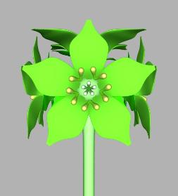
A
fictitious flower rendered from a 3D computer model that uses
mathematical functions. Click here to see how to make flower
models in Pov-Ray.
More
Flowers


![]()


Boraginaceae (Borage family)
![]()
![]()
Brassicaceae
(Cabbage family)
Rosaceae
(Rose family)
![]()
Caryophyllaceae (Campion or pink family)
![]()
Adoxacea (Moschatel)
![]()
![]()
![]()
Habitats

Salt marsh flowers (Chenopodiaceae)
Woodland flowers
Roadside
flowers
Meadow
flowers
Ponds
Even
More Flowers Info
Memorable
Flowers
- quotes about flowers
Yellow
Rose Flower
- meaning of flowers
Avas Flowers - floral arrangements and
bouquets
Freesia Flowers - greenhouse flower
arrangements
Hiking Flowers - flowers and plants to
see while hiking
Home Garden Flowers - flower tips for home
gardens

Article
last updated:
13/12/2014
23 August 2015
30 April 2016
26 June 2020
| |
|
|
|
|
|
|
|